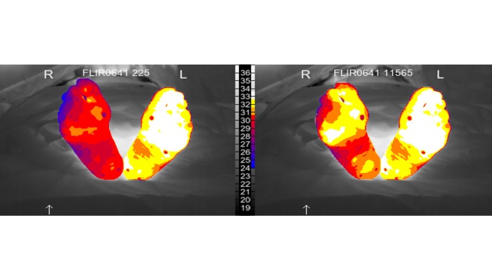
The technology, developed by Sheffield Hallam University, in collaboration with Sheffield Vascular Institute (SVI) at Sheffield Teaching Hospitals NHS Foundation Trust, has been trialled during treatment for patients with chronic limb-threatening ischaemia (CLTI), an advanced stage of peripheral arterial disease (PAD).
Reported in Frontiers in Medical Technology, Perfusion Right Now (PRN), uses infrared thermography and mapping, to monitor and track the effectiveness of minimally invasive (endovascular) procedures - such as angioplasty and stenting - to improve blood flow into the skin tissues of an affected limb. An outcome necessary for effective wound healing to take place in those patients with diabetic foot ulcers, or those at risk of losing a limb due to poor circulation.
PAD causes the arteries that supply blood to the limbs to become narrowed or blocked resulting in poor circulation, most commonly, to the lower leg and foot. Treatment includes medication and lifestyle changes, but severe cases can lead to chronic pain, non-healing wounds like ulcers, infections and even the loss of a limb. At this stage of the disease, known as CLTI, treatment is needed to reopen affected arteries and save the limb from amputation.
Despite endovascular procedures for CLTI having good technical success in restoring blood flow to the main affected arteries, the effectiveness of revascularisation to improve blood flow to the skin of the extremities is largely unknown. So clinical outcomes for patients following treatment can still vary considerably, putting them at risk of further interventions, or potentially a loss of their limb.
The development of PRN gives clinicians the imaging and analysis software tools to inform them about blood circulation ‘beyond’ the treated arteries for the first time. During the procedure they can observe if treatment is improving blood flow to the tissues of affected areas of a limb on a console and decide whether the procedure is likely to be successful.
The team behind the technology believe the results of the trial are the first to describe a new machine learning-assisted, non-invasive imaging and analysis system, designed to evaluate real-time tissue responses during procedures. They also highlight that PRN has potential for wider applications across medical and surgical specialities where understanding how effectively skin perfusion, or circulation into the tissues of the affected limb, is important.
Professor Charmaine Childs, from Sheffield Hallam University’s School of Health and Social Care, led on the development of PRN. She said: “Like many MedTech developments, PRN has had a long journey; from early testing and development for prediction of surgical wound infection to applications in the treatment of patients with narrowed or blocked arteries. The common thread here is the problem of insufficient perfusion of blood through the tiny vessels of the skin. This technology will be particularly valuable for treating patients with chronic diabetic foot disease, who have non-healing ulcers. At the end of the day, wounds need a good blood supply if they have any chance to heal.
“Our novel technology does not touch the body; it is relatively inexpensive and can be used continuously throughout the procedure. When used alongside current imaging methods, it will give the clinician a number representing the percentage improvement in skin foot perfusion from start to end of treatment to help understand whether healing is likely to be successful.”
As the technology is not reliant on visible light, PRN will potentially help reduce healthcare inequalities in effective skin assessment for people with a wide range of skin tones. So, to underpin their research, the team worked closely with the National Institute for Health and Care Research Doncaster Health Determinants Research Collaboration (HDRC), who engaged community leaders to reach demographic groups underrepresented in research, and gather inclusive data to ensure the technology would be effective for all patients.
Kevin Doyle, from the School of Health and Social Care at Sheffield Hallam, said: “Seeing the imaging analysis running live on the console in the angiography suite has been a real milestone, bringing our research closer to something clinicians can use in their practice. to assess foot skin responses to treatment.”
Professor Stephen Goode, Honorary Professor at Sheffield Hallam University and Consultant Vascular Interventional Radiologist at Sheffield Teaching Hospitals NHS Foundation Trust, said: “I regularly treat patients from around South Yorkshire with advanced PAD and CLTI who are facing the very real prospect of losing a limb, the difference between successful treatment and failure can ultimately mean the difference between keeping or losing a leg.
“Although we can successfully reopen blocked arteries using minimally invasive techniques, we cannot always be certain that enough blood is reaching the tissues to allow healing. That uncertainty is one of the greatest challenges in treating these patients.
“This new technology has the potential to fundamentally change that. For the first time, we are exploring the ability to monitor changes in tissue perfusion continuously during an intervention, giving us information that conventional angiography simply cannot provide. Ultimately, our goal is not just to open an artery - it is to heal wounds, relieve pain and save limbs. This research represents an exciting step towards making those outcomes more predictable for our patients.”
Professor of Machine Learning, Alex Shenfield, from Sheffield Hallam University, said: “This project blends cutting edge machine learning technologies with underpinning clinical science to deliver positive outcomes for patients both regionally and nationally. The outcomes demonstrated so far are a wonderful example of cross-disciplinary applied collaboration at Sheffield Hallam University, involving researchers across both the Colleges of Health and Wellbeing and Business, Technology and Engineering.”
Following the trial, the team are now conducting further evaluation of PRN, working to refine the proof-of-concept technology for clinicians, and are open to working with business and software partners to commercialise the technology.