Medical Image Processing and Visualisation
MRI, CT and Ultrasound Image Segmentation Arul N Selvan
A software for automatically segmenting dissimilar regions in medical images acquired with magnetic resonance imaging, computed tomography or ultrasonography has been developed and implemented. The software can be used to highlight abnormal regions in medical images.
The algorithm operates in unsupervised mode hence does not require any parametrisation. This allows the physician to fully focus on the task of identifying diseased areas.
The software is currently being evaluated and verified by a number of sonologists and radiologists.
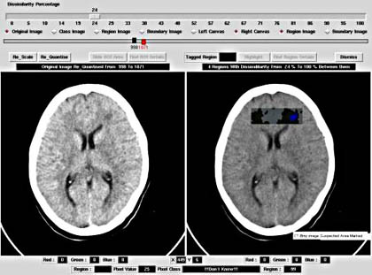
'The program has delineated the diseased area very clearly with a unique colour. The other part of the image with the same colour outside the diseased part of the image, can be differentiated by the physician as the non-diseased area as it is in the border of the uterus.
 Looking at the original image only an expert sonologist will be able to pick up this abnormality. Therefore this program will be surely useful to differentiate in case of borderline pathology where the sonologist will not be very sure of the diseased area.
The separate region within the diseased area gives additional information that, that area has got the core of the disease, and it is spreading out from that location'
- comment from a sonologist
Interactive Iso-Surface Visualisation of CT-data
In this project VTK and Tcl/Tk is used to implement visualisation software for 3-D data. One such example is the use of polygons generated using the marching cubes algorithm which is able to approximate iso-surfaces. (Note, that in the United States this idea is covered by a software-patent).
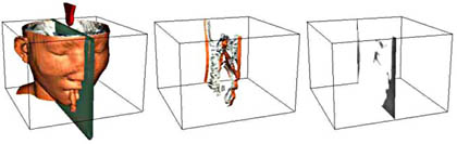
Left to right - two iso-surfaces, small slice through iso-surface-objects, density image of slice
Using an input-device with 6-DOF, it is possible to display segmented data in real-time, corresponding to the real-world position designated by the input-device.
3D Reconstruction and Visualisation of Breast Phantom internals using 2-D ultrasound image slices
This project involves the development of a calibration system for medical ultrasound machines.
Images captured at different focus levels are then related to the maximum depth. The project has implemented an untracked method of scanning breast phantoms in a systematic way.
Various preprocessing filters are applied to the acquired 2-D ultrasound images to facilitate 3-D reconstruction using the open source Visualization Tool Kit (VTK). (Trushal Kokate. Project supervisors: Arul Selvan and Bala Amavasai)
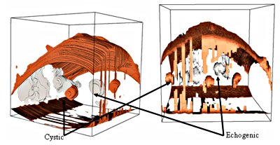
Different views of the 3D visualization produced using VTK for longitudinal plane.