Fingerprints have been the most powerful means of biometric identification for over a century. However, the process has remained largely unchanged in scope as only the physical characteristics of the ridge pattern are exploited to compare a crime scene mark with a fingerprint record, a process that leads to suspect identification if a match is found. Nonetheless, current fingerprinting may fail as in the case of smudged, partial and/or overlapped marks, or no fingerprint record.
An emerging and high TRL technology
In 2008, the Fingerprint Research Group (FRG) at Sheffield Hallam
University led by Professor Simona Francese, initiated the development of
Matrix Assisted Laser Desorption Ionisation Mass Spectrometry Profiling/Imaging
(MALDI MSP/I) based methods for the chemical profiling/imaging of fingermarks
(molecular fingerprinting), as well as their translation in an operational
environment.
Molecular fingerprinting enhances the forensic value of the fingermark
evidence by detecting and visualising the molecules that can be found within,
including contaminants, naturally occurring and introduced into the body.
This capability enables a novel way to conduct criminal profiling and to
inform crime scene management, narrow down the pool of suspects and speed up
investigations thanks to the provision of lifestyle and personal information as
well as chemical intelligence informing the dynamics and or the nature of the
crime.
The FRG pursued the
recovery of personal and lifestyle information around the finger mark's owner
and/or on their actions prior to or during committing the crime, enabling:
• Determination of the sex of the suspect from their fingermark peptidic/protein composition.
• Recovery of intelligence on the use of condoms, enabling associative/probative evidence in the context of crimes of sexual nature.
• Detection and mapping of drugs including date rape drugs.
• Specific and multi-informative detection of blood in marks/stains, compatible with CSI techniques.
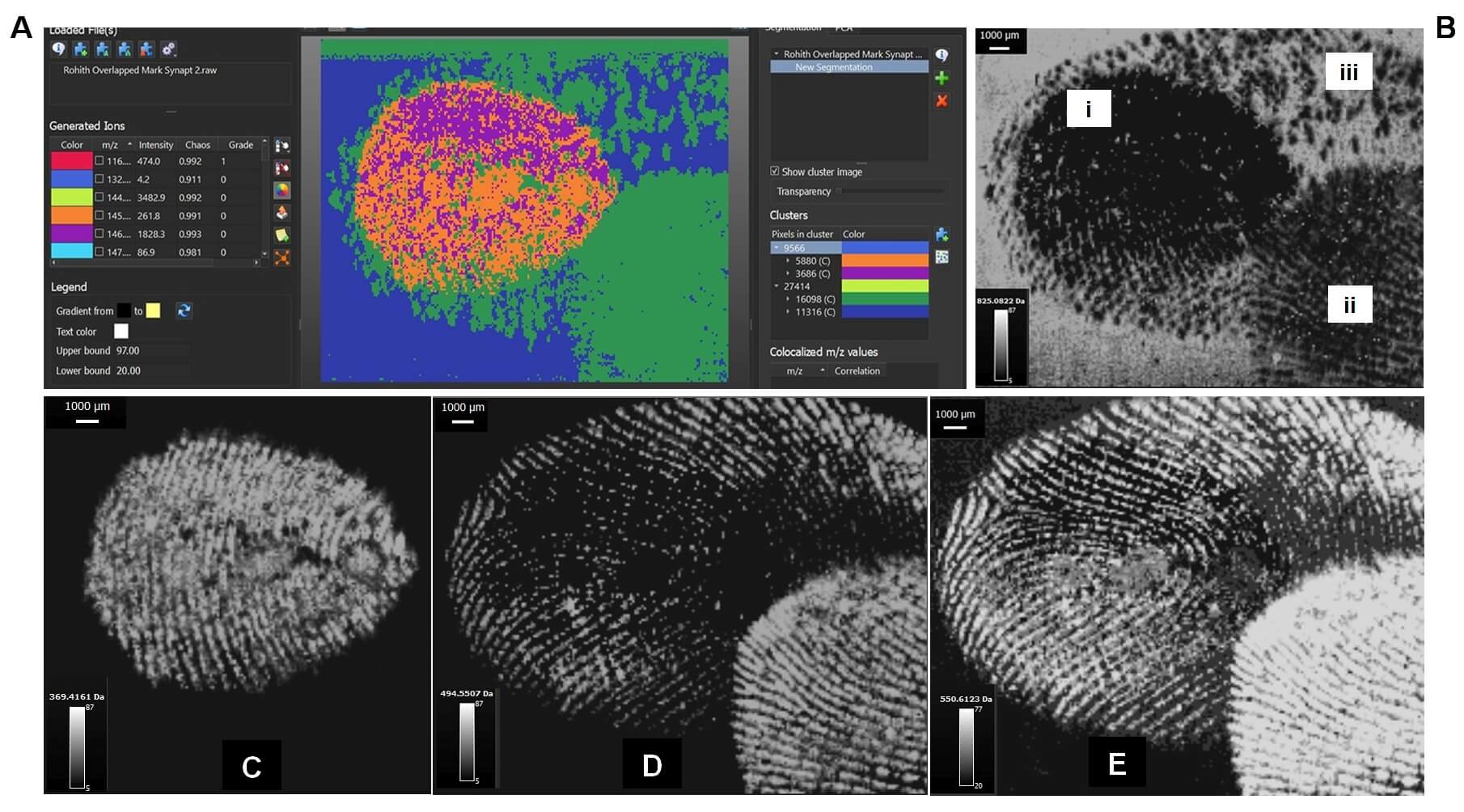
The ridge pattern can be reconstructed through visualising the molecules
that are detected while preserving the integrity of the evidence (Figure 1).