Using mass spectrometry and artificial skin to analyse skincare products

Get in touch

Contact SMIC to discuss facilities and partnerships.

Email SMIC

Using mass spectrometry and artificial skin to analyse skincare products

Skincare is big business, worth upwards of £2bn in the UK alone. Labskin UK and Sheffield Hallam are helping cosmetic and pharmaceutical companies find a competitive edge by using mass spectrometry and a market-leading artificial skin model to analyse product performance.

Beneath the surface

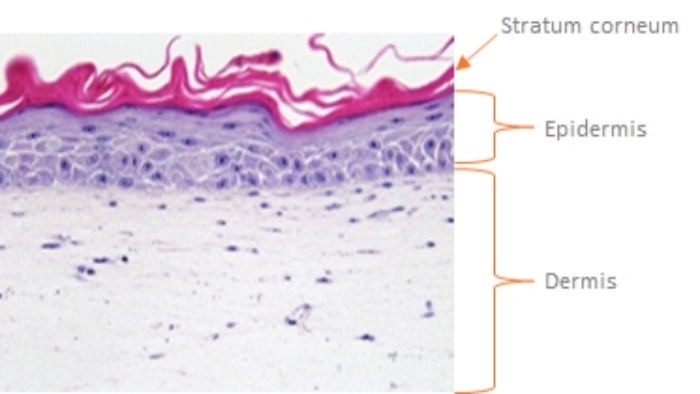
Skin is an incredible biological design. The largest organ in the human body, it provides a natural barrier to toxic agents, microorganisms and harmful radiation, while allowing essential substances like water and electrolytes to flow freely between cells. 

Cosmetics are designed to remain on the surface or sit in the outermost layers of the skin, which is known as topical delivery. While some pharmaceuticals are intended to pass through the skin into circulation, known as transdermal delivery. 

How they react with the skin depends on the physio-chemical properties of the active ingredient and the drug formulation1.

To see how these products actually perform, you need to look beneath the surface.

A living skin equivalent

Animal testing was banned under the 7th amendment to the European Union Cosmetic Directive in 2004. Since then, the market for artificial skin has surged, but due to its complex nature, skin isn’t easy to replicate. 

Labskin Ltd is a market leader, engineering 3D human skin models for ethical research and development. Because it mimics the skin’s biome, it’s an ideal model for testing cosmetic and skincare products. 

The full-thickness living skin equivalent (LSE) is grown with human skin cells on a scaffold material (Figure 1).

A section of Labskin, a living skin equivalent model of human skin produced by Labskin (UK) Ltd.
Figure 1: Haematoxylin & Eosin (H&E) stained image of a section of Labskin, a living skin equivalent model of human skin produced by Labskin (UK) Ltd York, UK (20x magnification).

Mass spectrometry imaging 

Since 2016, researchers at Labskin UK and Sheffield Hallam University have collaborated on skincare product studies. By using mass spectrometry imaging they’ve been able to analyse the absorption, metabolism and wound-healing properties of various applied skin products.

To do this, molecules are removed from the surface of the skin section as charged species (ions) and sorted by their mass to charge (m/z) ratio. 

Knowledge of the specific molecular mass of drug or cosmetic species allows their distribution in the skin section to be imaged.

For example, Figure 2 shows the distribution of the anti-fungal drug Terbinafine in a section of Labskin. These images were developed using the desorption electrospray ionisation mass spectrometry instrumentation housed in the Sheffield Multimodal Imaging Centre.

Here, the distribution of the active ingredient is clearly seen in the outermost (epidermal) layer of the skin model, verifying its use as a topical anti-fungal treatment as intended.

An Desorption Electrospray Mass Spectrometry Image of the distribution of the topical anti-fungal treatment terbinafine in a section of the living skin model Labskin showing that the drug is located in the upper layer of the skin model.
Figure 2: Desorption Electrospray Mass Spectrometry Image of the distribution of the topical anti-fungal treatment terbinafine (shown in green) in a section of the living skin model Labskin showing that the drug is located in the upper (epidermal) layer of the skin model.

The future of skincare

Sheffield Hallam and Labskin UK Ltd continue to collaborate on a number of projects with industry partners looking at the effects of pharmaceuticals and consumer health care products on healthy and diseased skin.

Footnotes

  1. Russo C et al Analytical Chem. 2018, 90, 16, 10031–10038

Get in touch

Contact SMIC to discuss facilities and partnerships.

Email SMIC