Using ultrasound to study how air pollution affects the human brain

Get in touch

Contact SMIC to discuss facilities and partnerships.

Email SMIC

Using ultrasound to study how air pollution affects the human brain

The brain consumes approximately 20% of the body’s total oxygen supply. Maintaining adequate flow of blood through the brain is essential to health. Using a transcranial doppler ultrasound (TCD), researchers at the Sheffield Multimodal Imaging Centre (SMIC) are studying how a common air pollutant, carbon monoxide, is affecting cerebral blood flow.

A non-invasive look into the brain

To study how this toxic gas affects human physiology, it is essential to be able to image cerebral blood flow. TCD is a non-invasive technique that uses ultrasound to measure the speed of blood flow through blood vessels in the brain. 

The most common blood vessel scanned is the middle cerebral artery (MCA), which can be found by placing the ultrasound probe on the temple and scanning at a depth of 35-55 mm. The MCA supplies major parts of the cortex and internal structures of the brain, and is the cerebral blood vessel most commonly affected by disease. 

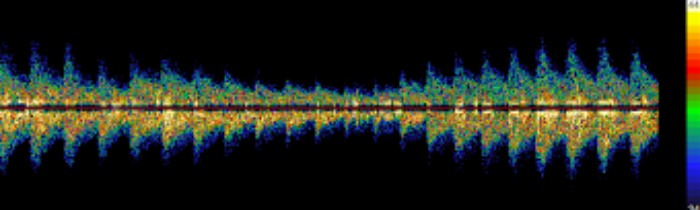
Often, the sonographer aims for the point where the middle and anterior cerebral (ACA) arteries originate from the internal carotid artery, which produces a characteristic waveform, seen in Figure 1.

Waveform of the  internal carotid artery bifurcation
Figure 1: Waveform of the ICA bifurcation. Signal from blood coming towards the probe (MCA) above the zero line, and blood going away from the probe (ACA) below the zero line.

The effect of a toxic gas

Researchers at SMIC, supported by the CO research trust, are using TCD to test blood flow following exposure to very low doses of carbon monoxide. 

Carbon monoxide, found in small amounts in air pollution, has the potential to alter blood flow. It may also change how cerebral blood flow is able to adjust to meet changes in oxygen demand. This is called cerebrovascular reactivity (CVR), and reduced CVR is considered a risk factor and marker for several diseases, such as stroke and vascular dementia. 

The research at SMIC shows that cerebral blood flow velocity begins to diminish even at carbon monoxide increases of 1 part per million in exhaled breath, and it is possible to observe how this happens beat-by-beat during inhalation of the gas due to the high temporal resolution of TCD (Figure 2). 

Middle cerebral artery (MCA) waveforms during CO inhalation
Figure 2: MCA waveforms at baseline and during CO inhalation; line is the maximum baseline blood flow velocity. Figure shows a progressive drop in velocity across 3 minutes of inhalation.

Researchers at SMIC are now testing to see how carbon monoxide alters CVR, and pairing TCD measurements with physiological measurements and other imaging modalities to get a full overview of what carbon monoxide does to our brains.

Bringing the laboratory to the field

TCD is a portable technique, and the equipment fits inside a small suitcase. It is therefore possible to use this methodology to test people in their homes, at work, and in areas of high air pollution, such as bus stations and car parks.

TCD can thus give us a unique insight into how environmental factors alter our brain blood flow, both in the laboratory setting and in people’s day-to-day lives.

Get in touch

Contact SMIC to discuss facilities and partnerships.

Email SMIC